| UniProt Accession Number | Reagent Type | Target Name / Protein Biomarker | Target Species | Host Organism | Isotype | Clonality | Vendor | Catalog Number | Conjugate | RRID | Availability | Method | Tissue Preservation | Target Tissue | Tissue State | Detergent | Antigen Retrieval Conditions | Dye Inactivation Conditions | Recommend | Agree | Disagree | Contributor | Notes |
|---|---|---|---|---|---|---|---|---|---|---|---|---|---|---|---|---|---|---|---|---|---|---|---|
| NA | Secondary Antibody | Rabbit IgG (H+L) | Mouse | Goat | IgG | Polyclonal | Thermo Fisher Scientific | A-21245 | AF647 | AB_2535813 | Stock | IBEX2D Manual | 1:4 Cytofix/Cytoperm Fixed Frozen | Lymph Node | NA | 0.3% Triton-X-100 | NA | 1 mg/ml LiBH4 15 minutes | Yes | 0000-0003-4379-8967 [2] | NA | 0000-0003-4379-8967 | |
| NA | Zenon Labeling Kit | Rabbit IgG (H+L) | Human | Goat | Fab2 | Polyclonal | Thermo Fisher Scientific | Z25308 | AF647 | NA | Stock | IBEX2D Manual | 1:4 Cytofix/Cytoperm Fixed Frozen | Lymph Node | NA | 0.3% Triton-X-100 | NA | 1 mg/ml LiBH4 15 minutes | Yes | 0000-0003-4379-8967 [2, 1] | NA | 0000-0003-4379-8967 | |
| NA | Secondary Antibody | Rabbit IgG (H+L) | Human | Donkey | IgG | Polyclonal | Thermo Fisher Scientific | A-31573 | AF647 | AB_2536183 | Stock | Cell DIVE-IBEX | FFPE | Tonsil | NA | 0.3% Triton-X-100 | pH 6 for 30 minutes ER1 (AR9961) and pH 9 for 30 minutes ER2 (AR9640) using the Leica Bond | 1 mg/ml LiBH4 15 minutes | Yes | 0000-0003-4379-8967 | NA | 0000-0003-4379-8967 | |
| NA | Secondary Antibody | Rabbit IgG (H+L) | Human | Donkey | IgG | Polyclonal | Thermo Fisher Scientific | A-31573 | AF647 | AB_2536183 | Stock | Cell DIVE-IBEX | FFPE | Lymph Node | Healthy | 0.3% Triton-X-100 | pH 6 for 30 minutes ER1 (AR9961) and pH 9 for 30 minutes ER2 (AR9640) using the Leica Bond | 1 mg/ml LiBH4 15 minutes | Yes | 0000-0003-4379-8967 [3, 4] | NA | 0000-0003-4379-8967 | 1 |
Publications
-
A. J. Radtke et al., “IBEX: an iterative immunolabeling and chemical bleaching method for high-content imaging of diverse tissues”, Nat. Protoc., 17(2):378-401, 2022, doi: 10.1038/s41596-021-00644-9.
A. J. Radtke et al., “Accompanying dataset for: IBEX: An iterative immunolabeling and chemical bleaching method for high-content imaging of diverse tissues”, doi: 10.5281/zenodo.5244550.
-
A. J. Radtke et al., “IBEX: A versatile multiplex optical imaging approach for deep phenotyping and spatial analysis of cells in complex tissues”, Proc Natl Acad Sci, 117(52):33455–33465, 2020, doi:10.1073/pnas.2018488117
-
A. J. Radtke et al., “Multi-omic profiling of follicular lymphoma reveals changes in tissue architecture and enhanced stromal remodeling in high-risk patients”, Cancer Cell, 42(3):444-463, 2024, doi: 10.1016/j.ccell.2024.02.001.
-
A. J. Radtke, “OMAP-18: Organ Mapping Antibody Panel (OMAP) for Multiplexed Antibody-Based Imaging of Human Lymph Node with Cell DIVE-IBEX”, Human Reference Atlas, 2024, doi: 10.48539/HBM356.HGHM.686.
Additional Notes
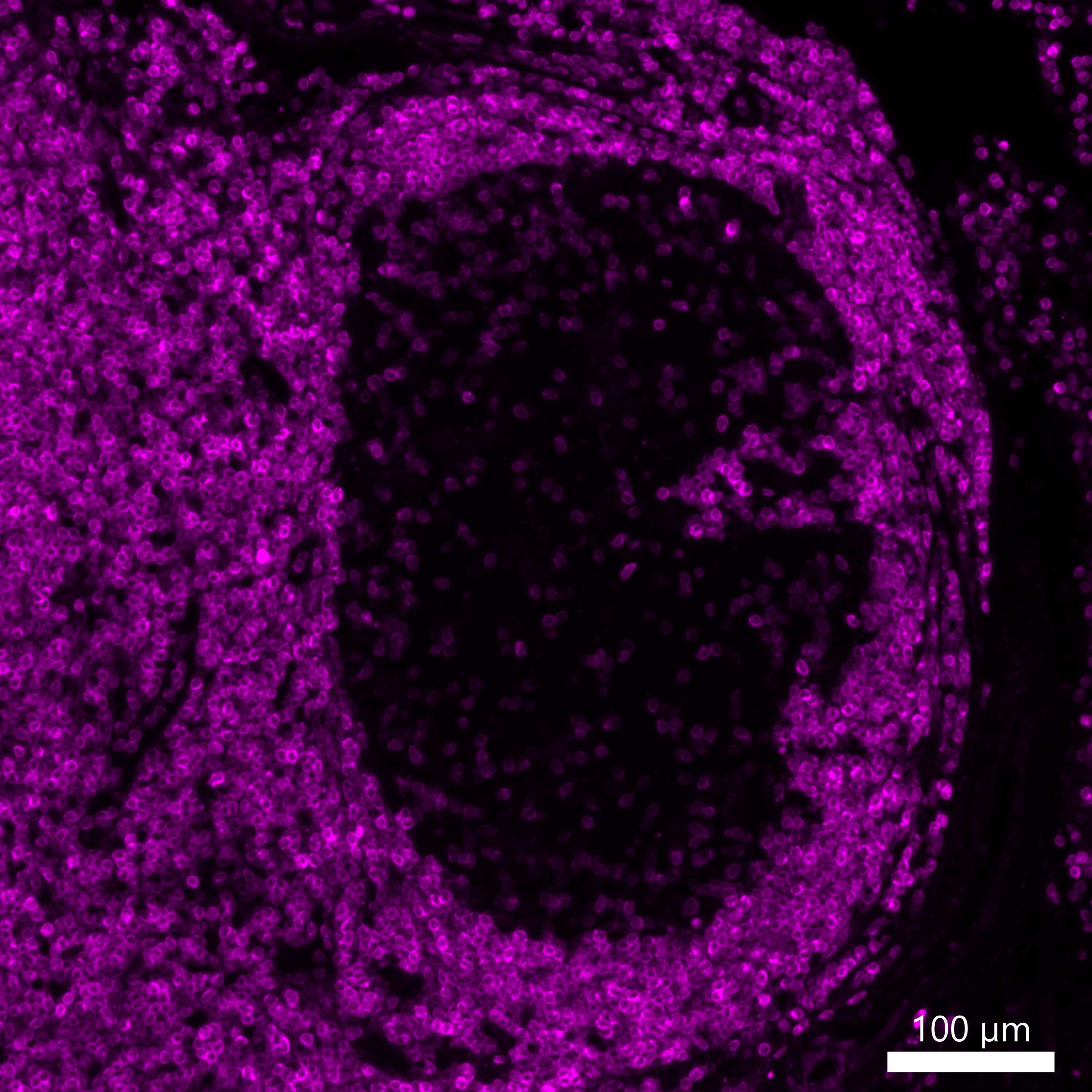
- Primary antibody (unconjugated BCL2) was used at a 1:500 dilution. Secondary antibody (Thermo Fisher Scientific A-31573) was used at 1:400 dilution.
| Human normal LN: BCL2 (magenta, catalog number ab236221 and A-31573) |
|---|
 |