| UniProt Accession Number | Reagent Type | Target Name / Protein Biomarker | Target Species | Host Organism | Isotype | Clonality | Vendor | Catalog Number | Conjugate | RRID | Availability | Method | Tissue Preservation | Target Tissue | Tissue State | Detergent | Antigen Retrieval Conditions | Dye Inactivation Conditions | Recommend | Agree | Disagree | Contributor | Notes |
|---|---|---|---|---|---|---|---|---|---|---|---|---|---|---|---|---|---|---|---|---|---|---|---|
| P51884 | Primary Antibody | Lumican | Human | Rabbit | IgG | EPR8898(2) | Abcam | ab252012 | PE | NA | Stock | Multiplexed 2D Imaging | FFPE | Lymph Node | Follicular Lymphoma | 0.3% Triton-X-100 | pH 6 for 30 minutes ER1 (AR9961) and pH 9 for 30 minutes ER2 (AR9640) using the Leica Bond | NA | No | 0000-0003-4379-8967 | NA | 0000-0003-4379-8967 | 1 |
Publications
Additional Notes
-
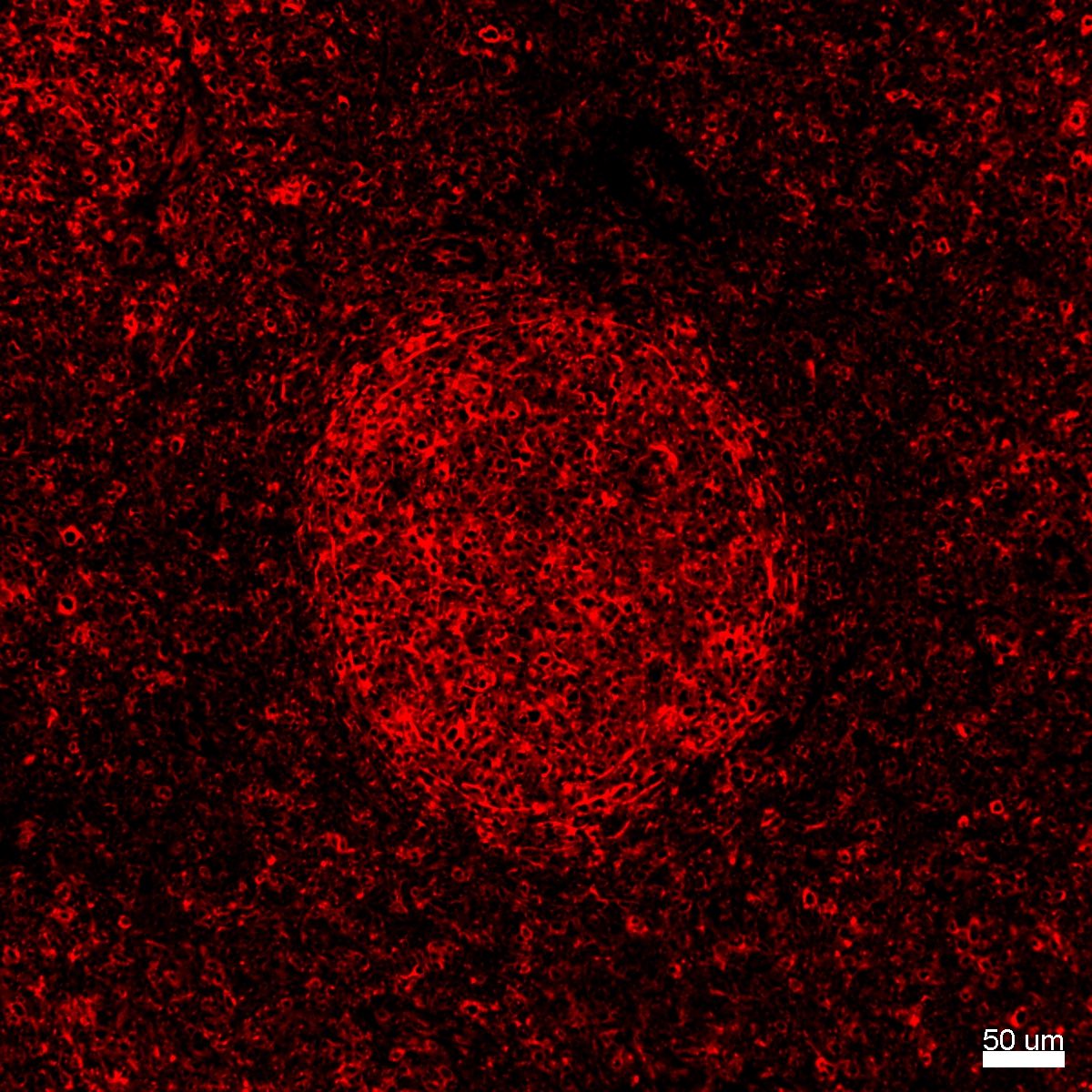
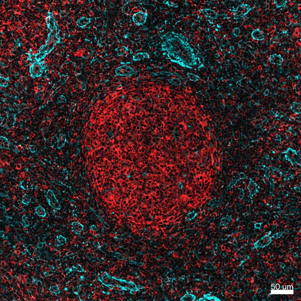
Beautiful staining pattern observed; however, antibody (Ab252012) appears to label B cells in the follicle (concentrated circle in center of image) and mesenchymal cells (based on appearance and location) outside of the follicle. Left image depicts lumican antibody (red, Ab252012) in follicular lymphoma FFPE sample prepared with dual antigen retrieval (pH 6 and 9). Right image demonstrates that Lumican PE (Ab252012, red) does not colocalize with a well characterized polyclonal antibody (Lumican R&D Systems AF2846, cyan) and secondary antibody (Thermo Fisher Scientific A-21084). Similar results, a follicular staining pattern, were observed in human FFPE tonsil samples.
Human follicular lymphoma FFPE : Lumican PE (red, ab252012 ) pH 6 and 9 Human follicular lymphoma FFPE : Lumican (cyan, AF2846) and Lumican PE (red, ab252012) pH 6 and 9