| UniProt Accession Number | Reagent Type | Target Name / Protein Biomarker | Target Species | Host Organism | Isotype | Clonality | Vendor | Catalog Number | Conjugate | RRID | Availability | Method | Tissue Preservation | Target Tissue | Tissue State | Detergent | Antigen Retrieval Conditions | Dye Inactivation Conditions | Recommend | Agree | Disagree | Contributor | Notes |
|---|---|---|---|---|---|---|---|---|---|---|---|---|---|---|---|---|---|---|---|---|---|---|---|
| NA | Primary Antibody | B220 | Mouse | Rat | IgG2b | RA3-6B2 | BD Biosciences | 571680 | RY775 | AB_3678612 | Stock | Multiplexed 2D Imaging | 1:4 Cytofix/Cytoperm Fixed Frozen | Lymph Node | NA | 1X BD PermWash Buffer | NA | NA | Yes | 0000-0002-6863-1461 | NA | 0000-0002-6863-1461 | 1 |
| NA | Primary Antibody | B220 | Mouse | Rat | IgG2b | RA3-6B2 | BD Biosciences | 571680 | RY775 | AB_3678612 | Stock | IBEX2D Manual | 1:4 Cytofix/Cytoperm Fixed Frozen | Lymph Node | NA | 1X BD PermWash Buffer | NA | 1 mg/ml LiBH4 15 minutes | Yes | 0000-0002-6863-1461 | NA | 0000-0002-6863-1461 | 2 |
| NA | Primary Antibody | B220 | Mouse | Rat | IgG2b | RA3-6B2 | BD Biosciences | 571680 | RY775 | AB_3678612 | Stock | Ce3D | 1:4 Cytofix/Cytoperm Fixed Frozen | Lymph Node | NA | 1X BD PermWash Buffer | NA | NA | No | 0000-0002-6863-1461 | NA | 0000-0002-6863-1461 | 3 |
Publications
Additional Notes
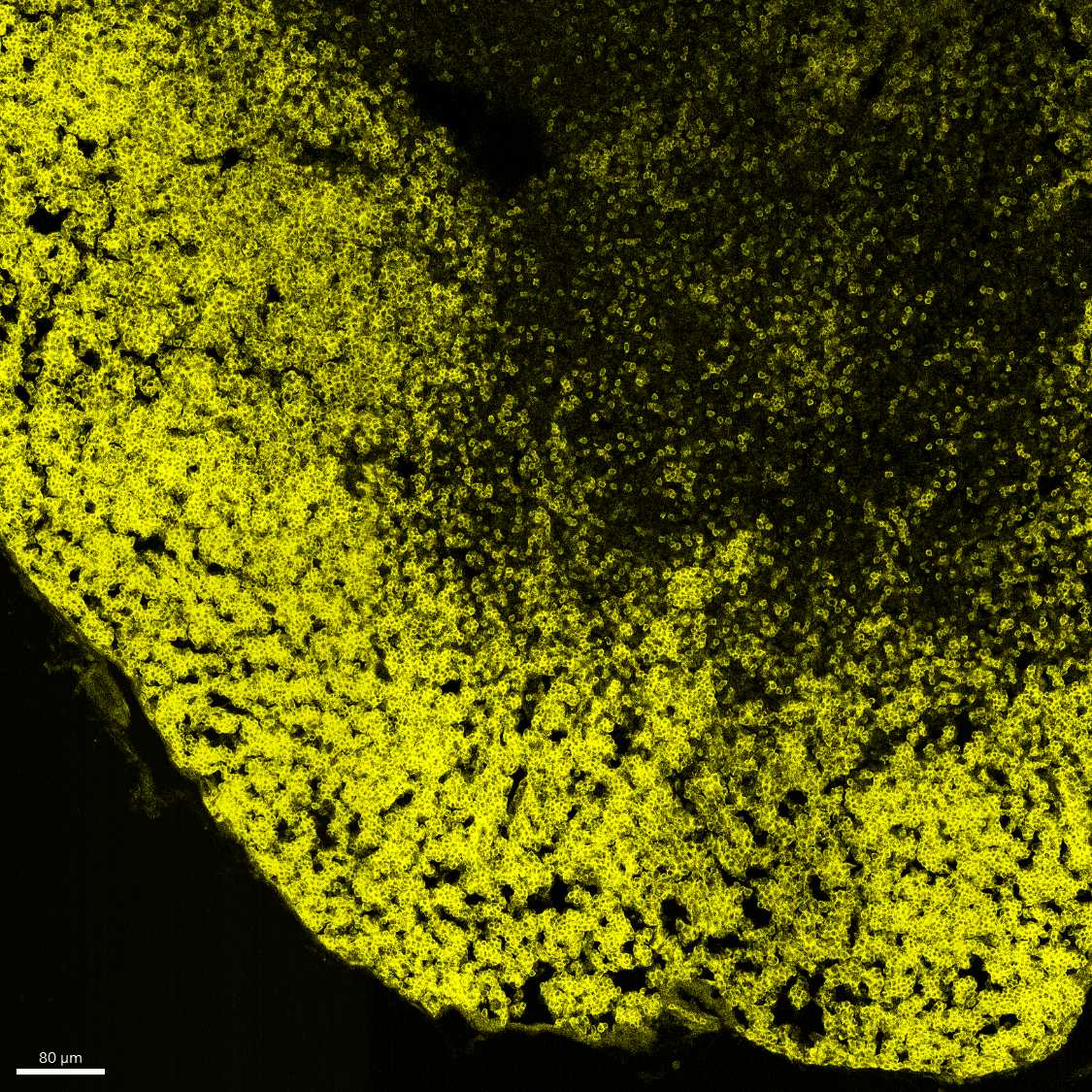
- Immunized draining lymph node. Stain was in 1X Perm Wash Buffer. Antibody concentration ~1/150, stained overnight at 4C.
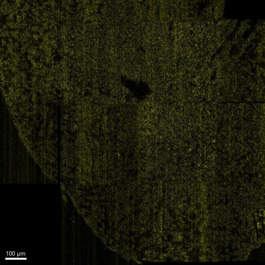
- Immunized draining lymph node. Stain was in 1X Perm Wash Buffer. Antibody concentration ~1/150, stained overnight at 4C. Dye inactivation was performed using 1mg/mL LiBH4 for 15 minutes.
- Immunized draining lymph node. Stain was in 1X Perm Wash Buffer. Antibody concentration 1/50, stained 3-4 days at RT on shaker. Did not post-fix. Cleared in Ce3D overnight. Not recommended due to poor penetration, but penetration might be improved at a higher conc (1/25).
| Immunized mouse lymph node |
|---|
 |
| Immunized mouse lymph node: dye-inactivated section imaged using pre-inactivation microscope settings (laser power, gain) but with signal maximally increased in Imaris to display the completeness of signal loss with dye inactivation. |
|---|
 |
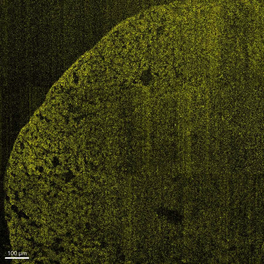
| Immunized mouse lymph node: dye-inactivated section imaged maximized microscope settings (laser power, gain) and optimal Imaris setting for visualization to evaluate potential residual non-inactivated signal which might interfere with subsequent imaging cycles, especially of dim markers. |
|---|
 |
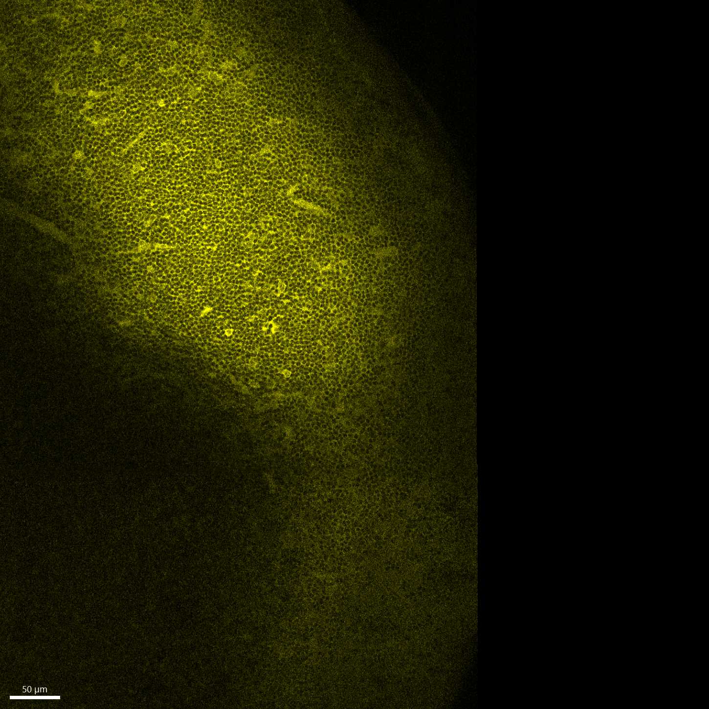
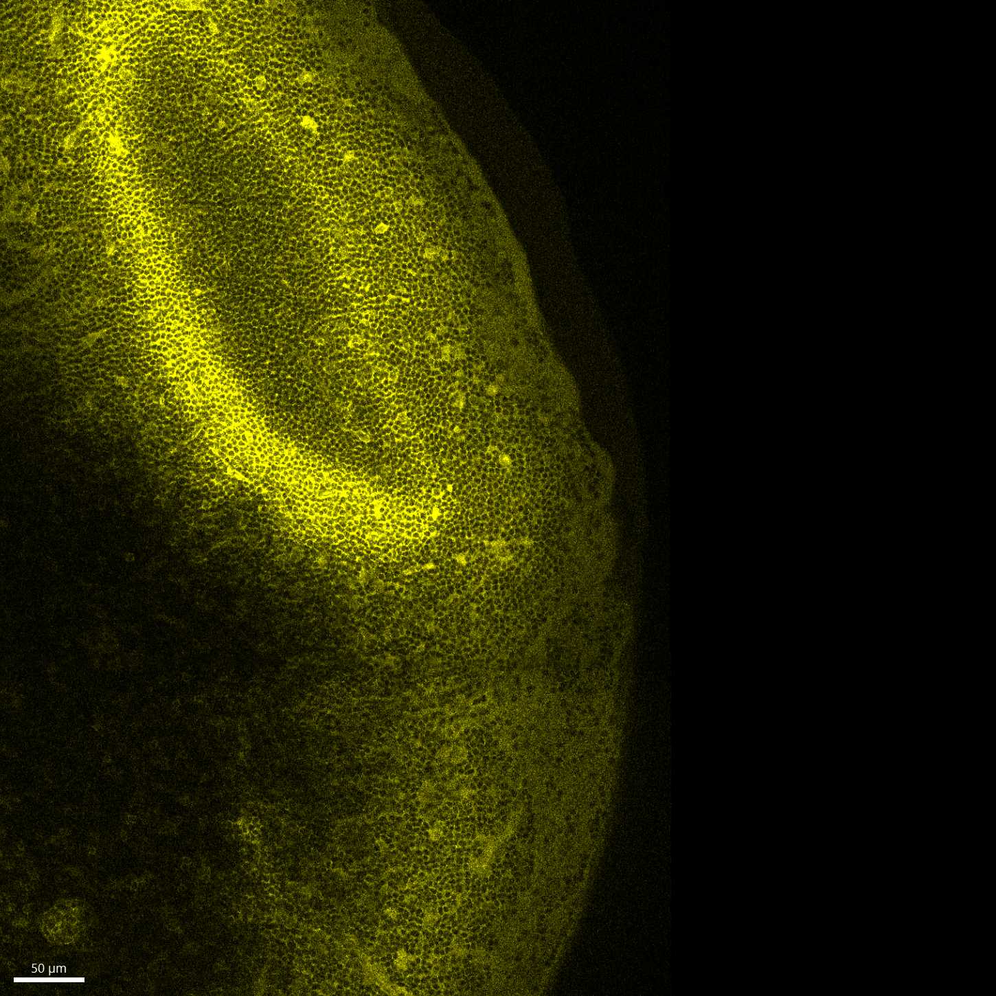
| Immunized mouse lymph node: 2D slice at the edge of a ~250um-thick 3D image |
|---|
 |
| Immunized mouse lymph node: 2D slice at the centre of a ~250um-thick 3D image |
|---|
 |